Cook-Calvete A, Delgado M, Jorquera S, Moreta S, Castro M, López J, Fernandez B, García J, Zaragoza C, Saura M. Endothelial ILK Deficiency Promotes Endothelial Activation and Cardiovascular Dysfunction via Receptor Interacting Protein Kinase-1 (RIPK1) Enriched-Extracellular Vesicle Signalling
J Extracell Vesicles. 2026
“Our study reveals how RIPK1-carrying extracellular vesicles can spread endothelial dysfunction and drive cardiovascular disease”. - Dr Alberto Cook Calvete & Dr Marta Saura Redondo
Summary:
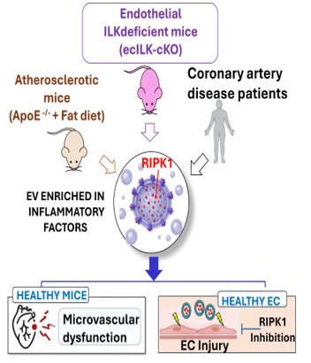
Integrin-linked kinase (ILK) maintains endothelial homeostasis by supporting endothelial nitric oxide synthase activity and restraining vascular inflammation. Although endothelial ILK loss is linked to vascular and cardiac disease, the mechanisms driving progression remain unclear. We hypothesized that extracellular vesicles (EVs) released by ILK-deficient endothelial cells propagate endothelial activation and cardiac dysfunction. In endothelial-specific ILK conditional knockout mice, ILK loss induced rapid endothelial activation, marked by increased iNOS, VCAM-1 and ICAM-1, followed by perivascular macrophage accumulation and chronic cardiac inflammation. In vitro, ILK-deficient endothelial cells exhibited barrier dysfunction, NF-κB activation and increased chemokine production. EVs derived from ILK-deficient cells were enriched in pro-inflammatory cargo, including receptor-interacting protein kinase 1 (RIPK1), and transferred the activation phenotype to naïve endothelial cells. RIPK1 inhibition in recipient cells or RIPK1 silencing in donor cells abolished EV-induced activation, thereby confirming a necessary role for RIPK1. Endothelial ILK-deficient mice also exhibited increased circulating EVs enriched in endothelial activation markers and RIPK1. Transfer of these EVs to wild type mice induced coronary endothelial activation, macrophage recruitment, microvascular remodelling, cardiac fibrosis, and ventricular dysfunction mimicking the phenotype of the donor mice. RIPK1-dependent inflammatory effects were similarly observed with EVs from atherosclerotic ApoE-/- mice and patients with coronary artery disease. These findings reveal an ILK-RIPK1 EV signalling axis that propagates vascular inflammation and promotes cardiac dysfunction, establishing a conserved pathogenic mechanism operative in experimental models and in human coronary artery atherosclerosis.
Why do you highligth this publication?
Our findings shift the concept of endothelial dysfunction from a local consequence of cardiovascular risk factors to a potentially self-propagating process. By identifying RIPK1-enriched EVs as vehicles that transmit endothelial injury from diseased to otherwise healthy cells, we provide a mechanism linking early vascular dysfunction to progressive coronary microvascular and cardiac remodeling. The presence of this pathway in atherosclerosis and coronary artery disease highlights its potential relevance to human cardiovascular disease and identifies the ILK-RIPK1-EV axis as a potential target for therapeutic intervention and EV-based biomarker development.
Publication commented by:
Dr Alberto Cook Calvete & Dr Marta Saura Redondo
CARDIOVASCULAR DISEASES group. IRYCIS