Microscopy, histology and comparative pathology

Leader

Ignacio Ruz Caracuel

Personnel

Silvia Sacristán (Histología)

Begoña Cuevas (Microscopía)

Contact

  • Equipment
    • CryoStar NX50 cryostat for frozen tissue samples.
    • Microm microtome model HM325 for paraffin-embedded tissue samples.
    • HybEZ II ACD hybridization oven.
    • Tissue microarrayers.
    • Extractor hood
    • Nikon ECLIPSE Ti confocal microscope with image acquisition system.
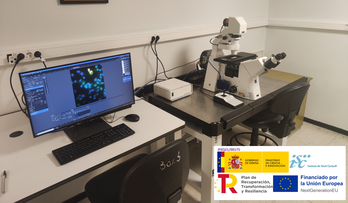
    • Zeiss Axio Observer 7 fluorescence inverted microscope.
    • ECLIPSE Ci fluorescence microscope equipped with Nikon DS-Ri1 camera.
    • Motic BA310 optical microscope equipped with MOTICAMS3 camera.
    • Image analysis workstation. NIS ELEMENTS software.
  • Noteworthy
    • Portfolio of services with basic histological techniques (in kerosene and frozen tissues), immunohistochemistry, immunofluorescence, aptahistochemistry, TUNEL and mRNA morphological detection systems.
    • Optical and fluorescence microscopy equipment, with confocal microscopy systems and fluorescence study of in vivo samples.
    • Advice on the interpretation of human histological sections and animal models by pathologists.
    • Acquisition of histological images and advice for quantification using free use software (Fiji, QuPath).
  • Services portfolio

    Microscopy


    **Consult fees with the unit

    Confocal Microscopy

    Confocal microscopy imaging of fixed samples (Multi-labeling imaging, multipoint colocalization experiments (X,Y,Z), FRAP)


    In vivo confocal microscopy

    Study of in vivo samples (cell cultures with fluorescent markers) along a time sequence using a cabinet cell incubation system with temperature, humidity, and gas control and perfusion system.


    Conventional and epifluorescence microscopy

    Acquisition of images with transmitted light (classical staining, immunohistochemistry) and from immunofluorescence using the digital imaging set with camera for photography. Obtaining large images formed by several individual fields for the study of areas of interest ("Stitching").


    Image processing and analysis

    Technical assistance in processing, quantifying and analyzing the information from the images obtained. An "off-line" workstation with specific software exclusively dedicated to image analysis is available.

    Histology


    Preparation of a paraffin block

    Includes dehydration of samples in successive alcohols of increasing concentration, rinsing in xylol and embedding in kerosene.

    Run 1 to 10 samples

    Cutting a paraffin block*.

    Cut sections of the paraffin-embedded sample, thickness between 5 and 20 microns, float the sections in a hot bath and mount the sections on slides. If the sections are to be stained with dyes we use albumin as an adhesive for the sections on the slides, but if they are to be used for immunohistochemistry we use special positively charged slides, which increases the cost. 


    Hematoxylin and Eosin Staining

    Harris hematoxylin (Papanicolaou's solution) and an alcoholic eosin solution should be used to stain the sections; the sections should be dehydrated and coverslipped, using Entellan as mounting medium. 


    Prepare a frozen block

    Impregnate histological specimens in 30% sucrose in phosphate-saline buffer, then coat with OCT for freezing in dry powdered ice, and store at -70°C.
     

    Cutting a frozen block*.

    Cutting of the frozen sample in a cryostat at a temperature of -20°C (may vary according to tissue hardness), obtaining sections of 10 microns thickness or greater, flotation of the sections in cold phosphate-saline buffer and mounting of the sections on slides. If the sections are to be stained with dyes, albumin will be used as an adhesive for the sections on the slides; if they are to be used for immunohistochemistry, special positively charged slides will be used, which increases the cost. 


    Immunofluorescence*.

    It includes the following successive treatments of the sections: unmasking of the antigen in a specific buffer at high temperature, blocking with non-immune serum and triton X-100, incubation in primary antibody and then detection of the antibody with a fluorochrome-conjugated secondary antibody. In the mounting medium used to cover the sections, Hoechst is included in order to visualize the nuclei of the cells present in the sections.
     

    Immunoperoxidase with signal amplification*.

    It includes the following successive treatments of the sections: inhibition of endogenous peroxidase with hydrogen peroxide, unmasking of the antigen in a specific buffer at high temperature, blocking with non-immune serum and triton X-100 and incubation in the primary antibody. A signal amplification technique consisting of successive incubations in a biotin-conjugated secondary antibody and in a peroxidase-conjugated streptoavidin solution and, finally, treatment with hydrogen peroxide and diaminobenzidine with which peroxidase reacts to give a brown product, will be used to detect the antibody. The nuclei of the cells are lightly stained with Harris hematoxylin. 


    * If the cuts give problems due to the consistency of the tissue or due to some problem in the preparation of the block, the cost will be increased according to the time of additional work to be carried out.

    * The primary antibody must be supplied by the client.